The Harris Lab has discovered that the geometry of the fruit fly embryo creates a crystal-like tissue structure. This revelation was published in the prestigious journal Development as “Confinement promotes nematic alignment of spindle-shaped cells during Drosophila embryogenesis“.
Professor Tony Harris’ laboratory studies the development of the Drosophila embryo to understand how cells become arranged before they become the organs in the body. As the embryo develops, a slab of cells called the germband folds over another group of cells called the amnioserosa.
Their research resolves a number of questions. Is the amnioserosa being held gently like you would hold a butterfly in your hand or squeezed like a stress ball? How is the amnisoerosa affected by its handling? What are the cellular structures that are involved?
Tirthankar Ray, PhD student in the Harris lab, studied whether these cells are under compression by cutting them with a laser. In the amnioserosa, the cells collapsed toward the cut site, indicating that the amnioserosa is under pressure from the germband like a stress ball.
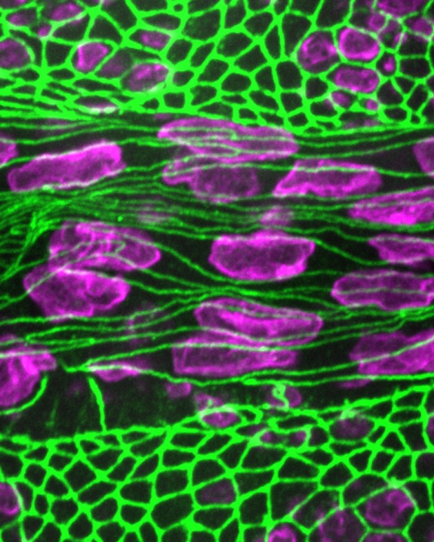
Ray, with the help of undergraduate researcher Damo Shi, went on to show that the germband pressure on the amnioserosa results in the cells becoming aligned as overlapping pointed cylinders, or spindles, like the crystals in a mineral.
Germband cell junctions are stabilized by actomyosin structures linked through the alpha-catenin protein. Ray determined that amnioserosa cells, by contrast, are kept in their spindle-like shapes by a scaffold within each cell composed of long microtubule strands.
Compared with germband cells, amnioserosa cells are less reliant on alpha-catenin to remain stably connected, and these cells interact with each other through a relatively unique type of cell-cell adhesion dependent on a protein called Baz/Par-3.
The alignment of many spindle-shaped cells is known as nematic ordering, a physical effect seen with rod-shaped objects from logs in rivers to liquid crystals. Reconstituted cytoskeletal networks and cultured cells were known to exhibit this behaviour, but Ray’s results show the relevance of nematic ordering to the development of animal embryos.
This study contributes a new element to embryo development by showing how organizational effects within a tissue can generate an array of spindle-shaped cells, and how these effects are combined with the physical influence of a tissue-tissue interaction that aligns the cellular array.