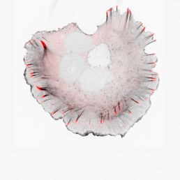
Prof Sergey Plotnikov studies how cell movement is guided by the stiffness of the tissue around the cell, and a new CIHR grant will allow his lab to continue pushing this work forward. Each day, cells in our body are continuously bombarded by a myriad of biological signals. As you are comfortably seated reading this article, these dynamic little beasts restlessly handle chemical signals while probing the mechanical properties of their surroundings. This active interaction between cell and environment is what allows cells to navigate the complex landscape they reside in.
The Plotnikov lab is particularly interested in understanding how the mechanical characteristics of tissue direct the movement of cells. Cell migration has been shown to underlie serious pathologies such as fibrosis and cancer metastasis. In each case, cells move directionally towards or along stiff areas of tissue.
This mesmerizing process can be observed in real time under a microscope. As cells crawl from one location to another, skeletal structures called actin filaments assemble and push the plasma membrane forward. Multi-protein structures called focal adhesions assemble in that area and act as little sticky feet to anchor the cell to the underlying surface. The coordinated assembly of focal adhesions at the cell front and disassembly at the cell back enables migration in a specific direction.
But how do cells measure the stiffness of the tissue around them to know where to go? The Plotnikov lab has revealed part of the decision process. In addition to their ability to anchor cell to surface, focal adhesions apply traction to the underlying surface and tug at it, just as we would repeatedly pull on an item to probe its texture. If the focal adhesions sense that the new area is stiff, they are reinforced and anchor more stably to the surface. If the new area is soft, the adhesions will not be as stable and will soon disassemble.
Equipped with several advanced microscopes, the Plotnikov lab uses an array of techniques to observe cell movement in 3D scaffolds, to visualize changes occurring in the cell’s skeletal structure, and to measure traction forces applied by each focal adhesion.
Having gathered this complex array of data, Plotnikov researchers augmented their observations with computational tools and genetic perturbations to find a specific ion channel located on the plasma membrane that is activated when focal adhesions pull on the surface below. They hypothesize that signaling pathways activated by this ion channel affect the stability of focal adhesions and the dynamics of the actin filaments that push the cell forward.
This prestigious CIHR grant will allow the Plotnikov lab to further decode the pushing and pulling forces mediating interactions between cells and their environment. Specifically, they aim to identify important players that allow cells to probe the mechanical properties of their surrounding and clarify the signaling pathways that enable migration towards stiff tissues.

