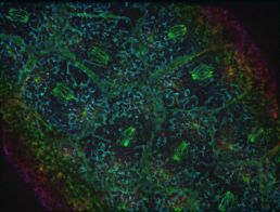
The Harris lab has revealed unexpected details about the puppetmasters of the cell in a new paper from Rebecca Tam in Journal of Cell Biology. Centrosomes are organelles that send microtubule threads throughout the cell. Molecular motors pull on these threads to move cellular structures around like puppets on strings.
Tam & Harris’ new study shows how centrosomes shape cortical caps on the surface of the Drosophila embryo by pulling on the cell surface. These caps delineate spaces that resolve into cells as the embryo develops.
Rebecca Tam was studying the cobblestoned pattern on the surface of the fruit fly embryo to understand how these rounded caps form on top of the nuclei within the embryo. In early stages of growth, the Drosophila embryo contains multiple capped nuclei in one large cell, a syncytium.
As the nucleus divides, astral microtubules emanate off centrosomes like rays of stars. Tam hypothesized that the astral microtubules pull the plasma membrane inward to initiate assembly of a cap.
The cell is packed with large structures between the centrosome and the embryo surface, which made it important to resolve how the centrosomes reach through this cluttered space. What Tam observed was that cap formation is dependent on multiple microtubules spreading out from the centrosome.
She found that the nascent cap is a local collection of plasma membrane folds with microtubules extending from their base toward the centrosome through the astral microtubule array.
In Tam’s microscope images, this looks like batter oozing through a sieve, with surface tension maintaining a flat area over the dividing nucleus. Tam shows that this pattern is dependent on pulling in of the plasma membrane by the centrosome as well as on global, actomyosin-based surface tension.
Arp2/3 actin network assembly on the plasma membrane infoldings promotes their unfolding into a cap, a process also promoted by exocytic vesicle association.
The comprehensive observations in “Centrosome-organized plasma membrane infoldings linked to growth of a cortical actin domain” provide a detailed view of pattern formation by a combination of local and global force-generating mechanisms. Centrosome-induced infoldings are shown to form a nexus of actin networks and a site of membrane growth for forming a new cortical domain.
Of all the graduate student publications in 2024, this paper earned Rebecca Tam the Dr. Christine Hone-Buske Scholarship for Outstanding Publication by a PhD Student! Congratulations, Rebecca!

