CSB Seminar: Improving functional axon regeneration in the injured nervous system
“Improving functional axon regeneration in the injured nervous system”
Alexandra Byrne, Assistant Professor
Department of Neurobiology, University of Massachusetts Medical School, MA, USA
Abstract:
To fully repair injured neurons, our nervous systems must both regenerate damaged axons and rebuild synapses with interacting cells, a process called functional axon regeneration. However, many injured axons, especially those in the central nervous system, actively inhibit functional axon regeneration, resulting in a permanent loss of motor and sensory function. Identifying and characterizing these molecular mechanisms that inhibit functional axon regeneration is critical to understanding how to repair the injured adult nervous system. To do so, we use a genetically tractable C. elegans model in which axon regeneration and degeneration can be studied in vivo with single axon resolution. I will present our recent findings of signal transduction pathways that function intrinsically and independently of one another to regulate axon regeneration, synapse reformation, and degeneration after injury. These include poly (ADP-ribosylation) and TIR-1/SARM signaling. Defining how these pathways regulate a neuron’s response to injury both contributes to strategies to improve functional axon regeneration after injury and adds to our understanding of the mechanisms that regulate post-developmental axon growth and synapse formation.
Friday, Jan 22nd, 2020 at 11:00 a.m.
https://utoronto.zoom.us/j/94049486250
Host: John Calarco (john.calarco@utoronto.ca)
CSB Seminar: Membrane remodeling in plant endosomal trafficking
“Membrane remodeling in plant endosomal trafficking”
Marisa Otegui, Professor
Departments of Botany and Genetics Laboratory of Cell and Molecular Biology U of Wisconsin-Madison
Abstract:
Cells rely on multiple degradative pathways to maintain protein and organelle homeostasis. One of these pathways, the endosomal route, requires extensive membrane remodeling and vesicular trafficking to sort and deliver proteins from the plasma membrane for degradation inside vacuoles. Whereas the molecular machinery in this pathway is largely conserved in all eukaryotes, plants have also evolved unique components accordingly to their particular cellular needs. I will discuss how ESCRT (Endosomal Sorting Complex Required for Transport) proteins mediate endosomal membrane remodeling during protein sorting in plants and how their functions control plant development and responses to environmental changes.
Friday, Jan 8th, 2021 at 11:00 a.m.
https://utoronto.zoom.us/j/94049486250
Host: Heather McFarlane: h.mcfarlane@utoronto.ca
CSB Seminar: Gene duplication, co-option and the evolution of cephalopod visual system complexity
“Gene duplication, co-option and the evolution of cephalopod visual system complexity”
Kristen Koenig, John Harvard Distinguished Fellow
Harvard University
Abstract:
The image forming eye is a classic model for the study of the evolution of biological complexity. The lens is a requisite innovation in all high-resolution, complex visual systems. The cephalopod has a highly acute visual system and is also a classic case of morphological convergence with the vertebrate eye. Both systems independently evolved a single-chambered eye with a cup shaped retina and a single refractive lens in the anterior. Almost nothing is known about the molecular-genetics of lens development in the cephalopod. The generation of new genetic material is considered a significant contributor to the evolution of biological novelty. We sought to understand if this mechanism may be contributing to cephalopod-specific visual system novelties. We identified a cephalopod-specific duplication of the transcription factor Sp6-9, with one paralog, DpSp6-9a, uniquely expressed in the lens-forming cells in the squid Doryteuthis pealeii. In addition, we find that DpSp6-9a is expressed during lens development in conjunction with a well-studied regulatory program canonically associated with proximal-distal patterning of animal appendages. To assess the homology of upstream regulation of these genes, we examined the role of Wnt signaling. We found that ectopically activating Wnt signaling leads to the loss of the lens and loss or decrease in limb-associated gene expression in the anterior segment. This work is a significant step forward in our understanding of the molecular basis of eye complexity in cephalopods, highlights the importance of transcription factor duplication, and sheds new light on the nature of phenotypes associated with co-opted canonical gene regulatory programs.
Friday, Dec 11th, 2020 at 11:00 a.m.
https://utoronto.zoom.us/j/94049486250
Host: John Calarco (john.calarco@utoronto.ca)
CSB Seminar: Early development and impacts on health and disease risk
“Early development and impacts on health and disease risk”
Deborah Sloboda, Professor
McMaster University
Abstract:
In recent decades the social and economic impacts of rising rates of chronic disease states, including obesity, diabetes, and cardiovascular disease, have prompted a global investigation into their causes as well as their consequences. Though initially considered to be largely determined by genetic and lifestyle factors, this paradigm would ultimately be insufficient to explain the continued propagation of non-communicable diseases. It is now established that perturbations during critical developmental windows result in (mal)adaptations that confer long-term disease risk, rather than health. Experimental and clinical studies have been essential in defining the nature and extent to which parental diet and metabolic status have on the developing fetus. In our work, we investigate how periconceptional nutritional adversity impacts maternal-fetal-placental relationships and impairs offspring reproductive and metabolic function. We show that maternal diet-induced obesity modifies maternal gut microbial communities, which may impact maternal metabolism through altered production of bacterial metabolites, including short-chain fatty acids (SCFAs) increasing intestinal permeability and altering maternal immune function. We show that paternal diet also participates in fetoplacental growth and development and influences placental development and in turn impairs offspring metabolic function. Regardless of the initiating lineage, an adverse in utero environment fuels changes in fetoplacental development and function, ultimately leading altered physiological function in the offspring long term.
Friday, Nov 27th, 2020 at 11:00 a.m.
https://utoronto.zoom.us/j/94049486250
Host: Jennifer Mitchell (ja.mitchell@utoronto.ca)
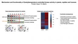
CSB Seminar: Mechanism and functionality of (body)temperature-controlled kinase activity in plants, reptiles and humans
“Mechanism and functionality of (body)temperature-controlled kinase activity in plants, reptiles and humans”
Florian Heyd, Professor
Department of Biology, Chemistry and Pharmacy, Freie Universität Berlin
Friday, Nov 13th, 2020 at 11:00 a.m.
https://utoronto.zoom.us/j/94049486250
Host: John Calarco (john.calarco@utoronto.ca)
CSB Seminar: Single-cell Studies of Phage Decision Making and Infection Dynamics
“Single-cell Studies of Phage Decision Making and Infection Dynamics”
Lanying Zeng, Associate Professor, Biochemistry & Biophysics
Texas A&M University
Abstract:
Bacterial viruses (bacteriophages) are the most abundant biological identities on earth. Quantitative studies of their infection cycles can elucidate the underlying mechanisms of the infection processes, shed important insight on how other viruses operate in higher organisms and help develop novel antibacterial strategies. In my talk, I will share two stories of different phages. In the first story, I will discuss our recent high-resolution studies of a paradigmatic system of cell-fate determination, the bacterium E. coli and its virus – phage lambda. Our studies suggest that individual phages vote for lysis or lysogeny and exhibit different interactions within the cell which include cooperation during lysognization, competition against each other during lysis, and confusion or coexistence between the two pathways. In addition, we found that phage DNAs establish separate subcellular compartments (‘ph(f)actories’) inside the cell which sustains heterogeneous viral development in single cells. In the second story, I will discuss our recent findings of ssRNA phages. We found that during the course of infection, ssRNA phage MS2 or Qbeta can detach the F-pilus of E. coli which is used for conjugation. This pilus-detachment feature can be thought of as a novel mechanism for superinfection exclusion and ssRNA phages can be engineered to remove retractile pili (virulence factors) thereby to disarm pathogens.
Friday, Nov 6th, 2020 at 11:00 a.m.
https://utoronto.zoom.us/j/94049486250
Host: Fernando Valencia (grad student host): fernando.valencia@mail.utoronto.ca
CSB Seminar: Building a SynBio toolbox to monitor and control plant hormone activity
“Building a SynBio toolbox to monitor and control plant hormone activity”
Anna Stepanova, Assistant Professor
North Carolina State University
Abstract:
Phytohormones are critical regulators of plant development and environmental responses. In the past three decades, the molecular pathways that govern hormone biosynthesis, signaling, and catabolism have been largely mapped out using a combination of genetics, molecular biology, biochemistry, and cell biology approaches. Despite the major progress, our ability to monitor and precisely control hormone action remains limited. With the development of inexpensive DNA synthesis technologies and the rise of synthetic biology as a new discipline at the intersection of molecular genetics and engineering, new molecular tools can now be built to enable hormone tracking and targeted hormone manipulation. We have generated a synthetic biology toolbox that allows rapid construction of multi-hormone transcriptional reporters. In addition, we are building CRISPR-based logic gate devices to confer novel, highly restricted patterns of expression to any genes of interest using a limited set of available native and synthetic drivers. The latter technology can be employed to tune the expression levels and subtract undesired domains of expression from existing drivers to precisely control output genes of interest, such as hormone biosynthesis, signaling, or catabolism genes, to regulate plant architecture, responses to stress, and other traits of interest. By combining multi-hormone reporters and genetic logic devices, we aim to shed fresh light on the mechanistic role of hormones in orchestrating plant development and stress physiology. That knowledge can then be relied upon to develop resilient next-generation crops.
Friday, Oct 16th, 2020 at 11:00 a.m.
https://utoronto.zoom.us/j/94049486250
Host: Shelley Lumba (shelley.lumba@utoronto.ca)
CSB Seminar: Biophysical mechanisms controlling tissue flows during development
“Biophysical mechanisms controlling tissue flows during development”
Karen Kasza, Assistant Professor
Columbia University
Abstract:
During embryonic development, groups of cells reorganize into functional tissues with complex form and structure. Tissue reorganization can be rapid and dramatic, often occurring through striking embryo-scale flows that are mediated by the coordinated actions of hundreds or thousands of cells. In Drosophila, cell rearrangements in the embryonic epithelium rapidly narrow and elongate the tissue, producing a tissue flow that doubles the length of the body axis in just 30 minutes. These types of conserved tissue movements can be driven by internal forces generated by the cells themselves or by external forces from neighboring tissue. While much is now known about the molecules involved in these cell and tissue movements, it is not yet clear how these molecules work together to coordinate cell behaviors, give rise to emergent tissue mechanics, and generate coherent flows at the tissue and embryo-scales. To gain mechanistic insight into this problem, my lab combines genetic and biophysical approaches with emerging optogenetic technologies for manipulating molecular and mechanical activities inside cells with high precision. I will discuss some of our recent findings on how cellular properties and mechanical forces are regulated in the Drosophila embryo to allow (or prevent) rapid cell rearrangements and tissue flows during specific events in embryonic development.
Friday, Oct 16th, 2020 at 11:00 a.m.
https://utoronto.zoom.us/j/94049486250
Host: Rodrigo Fernandez-Gonzalez rodrigo.fernandez.gonzalez@utoronto.ca
CSB Seminar: Cytoskeletal cross-talk with integrin adhesion complexes in cell morphogenesis
“Cytoskeletal cross-talk with integrin adhesion complexes in cell morphogenesis”
Alexander Bershadsky, Professor
Mechanobiology Institute, National University of Singapore
Abstract:
Cytoskeleton and cell-matrix adhesions are key elements determining cell morphogenesis. Here, two aspects of this topic will be discussed. (1) Microtubule-mediated crosstalk between adhesions and actomyosin. Transmembrane integrin adhesion receptors assemble into various types of actin cytoskeleton-associated structures, such as focal adhesions, fibrillar adhesions, and podosomes. These structures are controlled by microtubules via regulation of myosin-IIA-filaments assembly. Microtubules are coupled with integrin adhesions via KANK family proteins. This coupling controls release of guanine nucleotide exchange factor GEF-H1 from microtubules and its activation. GEF-H1 then activates Rho/Rho kinase, and thereby the assembly of myosin IIA filaments, which in turn remodel the adhesions. This mechanism appears to be universal for many cell types. (2) Emerging left-right asymmetry. Human fibroblasts confined to circular adhesive islands exhibit a chiral actin cytoskeleton swirling emerging due to unidirectional tilting of the focal adhesion-nucleated radial actin fibers. Analysis of molecular players by knocking down major actin-associated proteins and automated AI-based measurements of radial fiber tilting revealed a group of actin polymerization regulators required for the development of chirality. Depletion of other regulators reversed chirality direction. The confluent micro-cultures (of ~100 cells) confined to rectangular micropattern demonstrate a chiral cell alignment, an asymmetric tilt of average cell orientation relatively to the rectangle long axis. Analysis of more than 30 different knockdowns and pharmacological treatments revealed remarkable correlation between their effects on the chirality of individual cells and cell groups. Thus, actin-driven cell chirality could trigger the asymmetry in tissues and organs.
Friday, Oct 2nd, 2020 at 11:00 a.m.
https://utoronto.zoom.us/j/94049486250
CSB Seminar: On the front lines: How bacterial effectors at the host-pathogen interface govern host specificity
“On the front lines: How bacterial effectors at the host-pathogen interface govern host specificity”
Marcus Dillon, Assistant Professor
University of Toronto, Cell and Systems Biology
Abstract:
Bacterial pathogens pose a serious threat to global food security and human health. One critical virulence apparatus that pathogens deploy is the type III secretion system. The effectors that are delivered by this system mediate the outcomes of host-pathogen interactions because they can either promote pathogenesis on susceptible hosts or activate an effector triggered immune response on resistant hosts. Using the pangenome from nearly 500 strains of the agricultural pathogen Pseudomonas syringae, we conducted evolutionary genomic analyses to identify more than 14,000 effectors from 70 gene families. We then used a diverse representative collection of effectors to probe the effector triggered immunity landscape between P. syringae and the model plant Arabidopsis. We find that a shocking number of effectors elicit an immune response in Arabidopsis that reverses the outcome of the interaction from disease to resistance. These results illustrate that despite their widespread importance for pathogenesis, effectors may ultimately limit the host range of bacterial pathogens. My lab is now extending this pangenome approach to other bacterial phytopathogens, where we hope to identify host resistance genes that can provide stable resistance against a range of bacterial species.
Friday, Sept 25th, 2020 at 11:00 a.m.
https://utoronto.zoom.us/j/94049486250