Science is beautiful as well as practical
Our researchers have created stunning pictures that are on display throughout our facilities
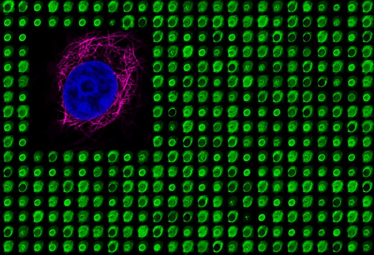
The emissions captured in these experiments can be given any colour, and overlaid with other selected colours
If you’re curious about the technical details of these images, check out our list below
An image of a larval zebrafish retina. Depicted in red and green are Muller glia, a cell responsible for monitoring the health of the retinal environment. The image is counterstained in blue to label all cell nuclei

Electron micrograph of the Golgi organelle

Output from the Bio-Analytic Resource for Plant Science depicting expression levels of the ABI3 gene and its relationships to other genes

Yellow microtubules in the pavement cells of the leaf

cells from the elongation zone of the Arabidopsis root

A panel of 4 Arabidopsis roots including root hairs

aniline blue-stained pollen tubes in the stigma (top) and along the style (bottom)

False coloured image of an electron micrograph of Drosophila (fruit fly) retina

A section through a wild type seed 14 days after fertilization by Jasmin Patel

Electron micrograph of a dechorionated Drosophila (fruit fly) embryo

Electron micrographs of two arabidopsis seed. left is a seed from a plant mutated in the FUS3 gene, right is an unmutated seed

Commissioned Art


