Microscopic detectors of viscosity – Plotnikov lab collaboration reveals how cells measure fluid thickness and move faster despite resistance
Researchers at University of Toronto and Johns Hopkins University have identified a specific cellular structure that allows cells to sense fluid thickness. In their recent Nature Physics article, the group explains how cells respond to thickened fluids and details the effect of viscosity on cell movement.
Cells in the human body are constantly moving. Cell movement not only allows the body to form organs and deploy immune cells to sites of infection, but also contributes to the progression of diseases such as fibrosis and cancer metastasis. Fast movement of cancer cells is particularly detrimental to cancer patients as malignant cells can quickly leave the original lesion and form new tumors in other parts of the body.
This new study reveals the mechanism that allows cancer cells to travel significantly faster in cancerous tissue. The fluid surrounding cancer cells, just like the mucus that surrounds lung epithelial cells in cystic fibrosis patients, is extremely viscous.
 Cell and Systems Biology Professor Sergey Plotnikov, who leads the Toronto team, was amazed how fast cells moved when they entered thick, mucus-like liquids. “Normally, we’re looking at slow, subtle changes under the microscope, but now we could see cells moving twice as fast in real time.”
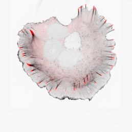
Researchers determined that the cell’s ruffling edges are key sensors of fluid viscosity. In normal liquids, ruffles at the cell edge continuously move up and down like flapping cuttlefish fins; however, when viscous fluid is added, the ruffles quickly flatten, spread out and latch on to the surface beneath them.
But how do the ruffles help speed cells up when cells should be slowing down due to resistance from the viscous fluid? To understand the driving force behind this counterintuitive observation, the group developed mathematical models and performed an in-depth analysis of cell mechanics. The combined efforts showed that it is in fact the resistance forces that flatten the ruffles and initiate a sequence of events that promote cell spreading and fast movement.
Specifically, flattening of the ruffles increases the contact time between the cell edge and the surrounding surfaces, which enables the cell to assemble more attachment sites to its surroundings. Long columns of actin, rod-like structures that give a cell its shape, grow from the attachment sites and push the edges of the cell outward. This increase in spreading is accompanied by an increase in cell-exerted forces that collectively push the cell forward.
The mechanism that helps cells measure the viscosity of surrounding fluids is not limited to cancer cells. Fibroblasts and macrophages also display similar ruffles and move faster in thick liquids.
“The fundamental science of the paper is of great interest,” says Prof Christopher McCulloch at the Faculty of Dentistry at University of Toronto, who further comments that the effect of viscosity on cells involved in blood and lymphatic diseases could be exciting to study.
The full details can be found in the article titled "Membrane ruffling is a mechanosensor of extracellular fluid viscosity" at Nature Physics.
Push and pull – the forces that earn Sergey Plotnikov a prestigious CIHR grant
Prof Sergey Plotnikov studies how cell movement is guided by the stiffness of the tissue around the cell, and a new CIHR grant will allow his lab to continue pushing this work forward. Each day, cells in our body are continuously bombarded by a myriad of biological signals. As you are comfortably seated reading this article, these dynamic little beasts restlessly handle chemical signals while probing the mechanical properties of their surroundings. This active interaction between cell and environment is what allows cells to navigate the complex landscape they reside in.
The Plotnikov lab is particularly interested in understanding how the mechanical characteristics of tissue direct the movement of cells. Cell migration has been shown to underlie serious pathologies such as fibrosis and cancer metastasis. In each case, cells move directionally towards or along stiff areas of tissue.
This mesmerizing process can be observed in real time under a microscope. As cells crawl from one location to another, skeletal structures called actin filaments assemble and push the plasma membrane forward. Multi-protein structures called focal adhesions assemble in that area and act as little sticky feet to anchor the cell to the underlying surface. The coordinated assembly of focal adhesions at the cell front and disassembly at the cell back enables migration in a specific direction.
But how do cells measure the stiffness of the tissue around them to know where to go? The Plotnikov lab has revealed part of the decision process. In addition to their ability to anchor cell to surface, focal adhesions apply traction to the underlying surface and tug at it, just as we would repeatedly pull on an item to probe its texture. If the focal adhesions sense that the new area is stiff, they are reinforced and anchor more stably to the surface. If the new area is soft, the adhesions will not be as stable and will soon disassemble.
Equipped with several advanced microscopes, the Plotnikov lab uses an array of techniques to observe cell movement in 3D scaffolds, to visualize changes occurring in the cell’s skeletal structure, and to measure traction forces applied by each focal adhesion.
Having gathered this complex array of data, Plotnikov researchers augmented their observations with computational tools and genetic perturbations to find a specific ion channel located on the plasma membrane that is activated when focal adhesions pull on the surface below. They hypothesize that signaling pathways activated by this ion channel affect the stability of focal adhesions and the dynamics of the actin filaments that push the cell forward.
This prestigious CIHR grant will allow the Plotnikov lab to further decode the pushing and pulling forces mediating interactions between cells and their environment. Specifically, they aim to identify important players that allow cells to probe the mechanical properties of their surrounding and clarify the signaling pathways that enable migration towards stiff tissues.