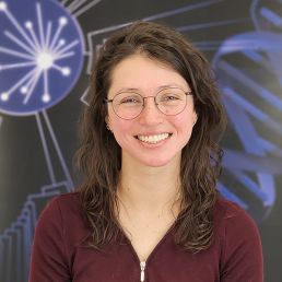
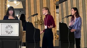
Tatiana Ruiz-Bedoya's passion for science yields Dr Christine Hone-Buske Scholarship
The Dr Christine Hone-Buske Scholarship honours an Outstanding Publication by a PhD Student in Cell & Systems Biology. This year’s award goes to Tatiana Ruiz-Bedoya for her work on “Cooperative virulence via the collective action of secreted pathogen effectors”.
Ruiz-Bedoya acquired an appreciation of natural diversity in her home city of Bucaramanga, Colombia and the nearby Chicamocha Canyon. From an early age, she sought to understand the natural world through science, proceeding to her current doctoral studies in Cell & Systems Biology.
In moving within the academic world through Bogotá, Uppsala, Munich and Boston prior to Toronto, Ruiz-Bedoya accumulated a prodigious amount of experience in genetics. Her interests range from identifying trafficked endangered animals through DNA to evolutionary genetics of phlox flower colour.
Ruiz-Bedoya was intrigued by research showing that the arms race between pathogens and host leads to rapid evolutionary changes. This passion led her to the Gutttman and Desveaux labs in Toronto, which study the host-pathogen response between plants and the bacteria Pseudomonas syringae.
Most plant pathology studies focus on individual strains found at high frequencies during outbreaks. But Ruiz-Bedoya notes that P syringae is an extremely diverse species complex with a life cycle that moves from clouds in the sky to soil surrounding potential plant hosts. Through this life cycle, there could be wide variation in virulence.
Ruiz-Bedoya and her lab colleagues reasoned that a focus on individual strains added bias to the way virulence was assessed. Within a carefully constructed community of non-virulent bacteria, they found that virulence could indeed emerge as a collective phenotype irreducible to its components.
Ruiz-Bedoya’s results form the very first proof-of-principle on the emergence of cooperation-driven virulence through the collective action of virulence factors. The findings of her award-winning research with Dr Pauline Wang, Prof David Guttman and Prof Darryl Desveaux was published in Nature Microbiology.
Ruiz-Bedoya attributes her success to stubbornness and to a determination instilled by her mother to go above and beyond. She is excited by science and her passion for research is something she emulates from seeing it in her many mentors. “Some of these qualities can be as helpful as they can be upsetting, especially when experiments don't work”, says Ruiz-Bedoya, “But you must be determined to find the balance within your life and I am very privileged that I get to do science for a living.”
With additional roles as a teaching assistant, communicator and friend, Tatiana reflects the spirit of Dr Christine Hone-Buske, who approached her work as a student, teaching assistant, and researcher at U of T with confidence, curiosity and drive.
Congratulations, Tatiana Ruiz-Bedoya, on your Dr. Christine Hone-Buske Scholarship for Outstanding Publication by a PhD Student!
Mitchell lab reveals redundant systems that maintain genome architecture
Redundancy ensures integrity of gene expression independent of genome architecture
Part of finding out how something works is trying to break it. Two recent papers from Professor Jennifer Mitchell’s lab try to break the folded structure in the genome that links the essential Sox2 gene with its far away regulators. But no matter how many elements they removed or added around the Sox2 region, this structure was maintained, revealing the depths of the redundant systems that protect genome integrity.
Sox2 is crucial for the identity of embryonic stem cells. Dr Ian Tobias, a postdoctoral fellow in the Mitchell lab in Cell & Systems Biology, notes that “It makes a lot of sense, from an evolutionary and developmental biology standpoint, that such a key regulator of cell fate wouldn't be so fragile as to have its regulatory control lost with the deletion of a small segment of DNA.”
Sox2 and its distant control region are bundled together
Inside our cells the genome consists of long ribbons of DNA that contain all the information to make every part of the body. In organs like lung or brain, the DNA is fenced off in different ways to turn on lung or brain-specific genes, while hiding genes that should not be active. The Sox2 gene region is open and active in embryonic stem cells but closed in most other cell types.
Active Sox2 in embryonic stem cells is isolated within a region that contains only the gene and its activators including the Sox2 Control Region (SCR). The SCR is over 100,000 base pairs away from Sox2, so this region is relatively long. Individual organised regions of the genome have been called topologically associated domains (TAD), and the Mitchell lab studies how these domains are established and maintained.
Tiegh Taylor, a PhD student in the Mitchell lab used gene editing to tease out the mechanisms that keep the Sox2-SCR gene domain intact. Working with colleagues in the Sexton lab in Strasbourg, France, their studies of the region around Sox2 challenged the dogma of how the winding ribbons of DNA in a cell are organized.
In large scale studies of the genome, the CTCF protein is almost always found at TAD boundaries, leading to the idea that CTCF-bound DNA acts as a fence that isolates DNA outside of the domain from DNA inside the domain. Separate transcription factor proteins are responsible for individual gene activation, bundling DNA within the TAD together.
CTCF is not required for Sox2 TAD maintenance
What the Mitchell and Sexton labs showed is that transcription factor proteins can still keep DNA organized in embryonic stem cells even in the absence of the CTCF fences.
How did they show this? The SCR is comprised of two clusters of transcription factor binding sites flanking a CTCF site. At the Sox2-SCR loop, transcription factor proteins interact to enhance gene activation, giving the SCR the designation of an “enhancer”.
When the researchers removed the transcription factor-binding clusters, Sox2 enhancement was lost, but the TAD bundle remained in place. Removing all of the SCR including the CTCF site loosened the bundle to interact with an adjacent TAD, so presumably CTCF is holding the TAD together.
Surprisingly, Taylor found that although this CTCF site was sufficient to fence in the TAD, it wasn’t necessary. When removed from the SCR, the TAD bundle was maintained, presumably due to the transcription factors binding to the enhancer surrounding the missing CTCF binding site. The strength of these transcription factors is so strong in fact, that they are able to ‘jump over’ an artificial CTCF fence which was later placed between Sox2 and the SCR.
Taylor notes that “It wasn’t clear what the finding was at first, as the data came in it seemed like nothing we could do would break this interaction. It wasn’t until we thought about it for a while that we realized that maybe it’s not that these various proteins are so defined in their roles of interaction vs enhancer, it’s more about this complex cooperativity between all these factors that makes this locus so robust. It really highlights just how complicated our genome is.”
This work was published in the journal Genes & Development as “Transcriptional regulation and chromatin architecture maintenance are decoupled functions at the Sox2 locus”
Transcription factors maintain Sox2 architecture, even as stem cells become neural progenitors in mice
Taylor’s colleague Dr Ian Tobias is studying what happens to enhancer activity when embryonic stem cells change into other cell types where Sox2 is active. “We're interested in the neural lineage because it's also highly dependent on the Sox2 protein; the model we use is neural progenitor cells (NPS). During the differentiation of embryonic stem cells to NPS, Sox2 interacts with a region that’s even further away than the SCR.”
Using techniques similar to Taylor’s approach, Tobias found that when the far away region is deleted, Sox2 doesn’t turn on in NPS, making this site a candidate Sox2 distal neural enhancer or DNE. At the same time, collaborators at the NIH in Maryland were looking at TADs in neurons and saw a clear domain linking the DNE to Sox2. When the NIH group blocked TAD formation in mice by inserting CTCF between the DNE and Sox2, interaction was reduced, but not eliminated, again showing how robust these interactions can be.
The result of this collaboration has been published in Nature Genetics as “Enhancer–promoter interactions can bypass CTCF-mediated boundaries and contribute to phenotypic robustness”.
New targets for understanding genome architecture and disease
The combined results in these two papers reveal that specific elements supplying enhancer function contribute to maintaining DNA structure. They consequently argue for looking beyond CTCF when looking to explain how DNA folds in the nucleus.
Professor Mitchell notes that “Now that we know these enhancer sequences are important for regulating Sox2 during development, our next steps are studying aberrant expression of Sox2 in disease.” Tobias and others in the Mitchell lab will be pursuing the role of Sox2 enhancers and their associated proteins in neurological disorders and other diseases.
Professor John Peever earns Distinguished Scientist Award from the Canadian Sleep Society
Professor John Peever is this year’s recipient of the Canadian Sleep Society's (CSS-SCS) Distinguished Scientist Award.
This award recognizes a clinician or scientist who has made significant national and international contributions to the field of sleep medicine and biology. The recipient is invited to give a Keynote address at the CSS-SCS National Conference, held this year on April 27-29 in Ottawa.
Research in Peever's lab is focused on identifying the brain circuits that control sleep and wakefulness, and how breakdown in these circuits contribute to disorders such as Parkinson’s disease and narcolepsy. He is also a strong advocate for promoting the awareness of sleep in health and disease.
Peever asserts that "It’s an honour to receive this recognition but all the credit goes to my outstanding team of trainees!" You can read more about Peever and his trainees' work in this story: "U of T neurobiologists discover switch that turns muscles on and off", and in Research 2 Reality's video profile: "What goes into a good night's sleep?"
Congratulations, Professor Peever!
Sustainability award to Chief Horticulturalist Bill Cole for greenhouse energy improvements
U of T's Chief Horticulturalist, Bill Cole, has led the change for energy efficiency in our plant growth facilities through relentless effort on lighting retrofits. Cole's work has earned him the Sustainable Action Award from Facilities and Services.
Plant science requires tightly controlled temperature and lighting, whether in a growth chamber or a greenhouse, but the power demands for these conditions can approach industrial levels. Earth Sciences Building greenhouses contain specimens representing hundreds of plants in specialized environments to mimic desert, tropical and temperate zones.
In 2021, Cole's team retrofitted one zone of the Earth Sciences Building greenhouses from old yellow sodium lamps to LED panels. This pilot project reduced energy consumption and lowered heat output. There was an added benefit of more blue and red wavelengths to enhance growth of research and teaching plants. The improved lighting will allow growth of light-hungry research plants from Brazil and South Africa.
Goldleaf Technologies has provided 200 Thrive LED Grow Lights to retrofit all 15 greenhouse zones with LED panels starting in January 2023. Goldleaf notes that their LED lights provide over 50,000 hours of light, losing only a fraction of intensity over their lifespan compared to obsolete lighting systems that require frequent repair.
The Greenhouse facility operation team has the experience to operate this specialty lighting and has developed a detailed schedule to upgrade them. The new LED panels will be installed over two months, gradually changing the yellow glow atop the Earth Sciences Building to a more even hue. This will reduce energy consumption by 33%, moving toward U of T's goal to be net Carbon positive by 2050.
Cole donated a portion of his Sustainable Action Award to Sustainability Network, whose mission is to strengthen environmental nonprofit leadership.
Congratulations, Bill Cole!
CSPB awards for CSB's dedicated plant scientists
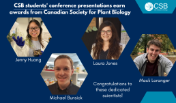
CSB students won multiple awards at the annual regional meeting of the Canadian Society for Plant Biology, held at University of Toronto Scarborough on December 3, 2022.
Jenny Huang (McFarlane lab) noted that "It was exciting to finally meet all the scientists I had only heard of by name before. The conference exposed me to a diverse range of research in the field of plant science which broadened my perspective on the kinds of projects that are available."
Huang earned her award for a talk on Sugar Code Interactors in N-glycan Quality Control. She says "I was not expecting to win an award, and I am thankful to have my work recognized". Lectins bind to N-glycosylated proteins, and Huang has identified candidate lectins in Arabidopsis with the goal of exploring their effect on glycoprotein trafficking and maturation.
Michael Bunsick (Lumba lab) was recognized for his talk on Identifying the Strigolactone Receptor in Fungi. Bunsick found that plant-derived strigolactone serves as an inter-kingdom signaling molecule which inhibits PHO84-dependent phosphate uptake in fungi.
Mack Loranger (Yoshioka lab) presented an award-winning poster describing how Diverse interactions between Solanum lycopersicum (tomato) and beneficial Canadian soil-borne bacteria regulate increased immunity.
Laura Jones (Braeutigam lab) says she was "thrilled to receive a poster award for my research on Responses of Balsam Poplar to Extreme Weather Events Simulating Late 21st Century Climate. It felt like a confirmation that people recognize the importance of studying tree physiology, particularly in the context of climate change, and find trees just as fascinating as I do!"
Congratulations to these dedicated scientists!
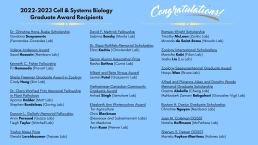
Congratulations to CSB's Graduate Student Award Recipients!
Congratulations to our Graduate Students who earned recognition for their accomplishments at our Graduate Student Awards on December 19th, 2022!
Valerie Anderson Graduate Fellowship
- Awarded for academic merit to an outstanding student in any subdiscipline of plant biology.
Recipient: Saad Hussain (Nambara Lab)
Kenneth C. Fisher Fellowship
- Awarded in recognition of a student who maintains a high standard of academic and research achievement, balanced with outstanding extra-curricular contributions to their department.
Recipient: PJ Gamueda (Provart Lab)
Sheila Freeman Graduate Award in Zoology
- Awarded to an incoming or in-progress graduate student focusing their studies in animal biology.
Recipient: Cindy Hong (Kim Lab)
Dr. Clara Winifred Fritz Memorial Fellowship in Plant Pathology
- Awarded to students studying in the area of plant pathology demonstrating academic excellence.
Recipients: Aparna Haldar (Mott Lab)
Stephen Bordeleau (Goring Lab)
Duncan L. Gellatly Memorial Fellowship
- Awarded each year to one or two graduate students demonstrating excellence in Virology and/or Molecular Biology research
Recipients: Arvin Persaud (Guzzo Lab)
Tiegh Taylor (Mitchell Lab)
Yoshio Masui Prize in Developmental, Molecular or Cellular Biology
- Awarded to a master’s or doctoral student in the Department on the basis of academic merit.
Recipient: Gerald Lerchbaumer (Tepass Lab)
David F. Mettrick Fellowship
- Awarded to a graduate student in CSB engaged in any aspect of zoological research.
Recipient: Sabrina Barsky (Monks Lab)
Dr. Klaus Rothfels Memorial Scholarship
- Scholarship awarded on the basis of academic standing.
Recipient: Elina Kadriu (Christendat Lab)
Senior Alumni Association Prize in Cell & Systems Biology
- Awarded to a student in the department based on academic merit.
Recipient: Nasha Sethna (Currie Lab)
Hilbert and Reta Straus Award
- Awarded to a full-time graduate student who has demonstrated high research achievement in the fields of plant molecular or cellular biology.
Recipient: Jasmin Patel (Gazzarrini Lab)
Vietnamese-Canadian Community Graduate Award in Zoology
- Awarded to a master’s or doctoral student studying animal biology based on academic merit and who exhibits research potential, excellent communication skills and leadership.
Recipient: Anhad Singh (Senatore Lab)
Elizabeth Ann Wintercorbyn Award
- This first award is made to a student engaged in research work which is likely to prove beneficial to medicine.
Recipient: Ryan Ruan (Peever Lab)
Elizabeth Ann Wintercorbyn Award
- The second half of the award goes to a student engaged in research work which is likely to prove beneficial to agriculture.
Recipient: Chris Blackman (Desveaux and Subramaniam Labs)
Ramsay Wright Scholarship in Cell and Systems Biology
- Awarded to CSB students engaged in research in zoology.
Recipients: Timothy McLean (Zovkic Lab)
Miranda de Saint-Rome (Woodin Lab)
Zoology International Scholarship
- This is the first of two awards for international students demonstrating high academic performance.
Recipient: Manisha Kabi (Filion Lab)
Zoology International Scholarship
- This is the second award, again, going to international students demonstrating high academic performance.
Recipient: Jiashu Liu (Liu Lab)
Zoology Sesquicentennial Graduate Award
- Awarded to a graduate student enrolled in full-time studies in CSB, on the basis of academic merit.
Recipient: Haoyu Wan (Bruce Lab)
Alfred and Florence Aiken and Dorothy Woods Memorial Graduate Scholarship in Cell and Systems Biology
- Awarded based on academic merit.
Recipients: Osama Abdalla (Cheng Lab)
Mahbobeh Zamani Babgohari (Gonzales-Vigil Lab)
Rustom H. Dastur Graduate Scholarship in Cell and Systems Biology
- Awarded to a graduate student studying plant sciences, on the basis of academic merit.
Recipient: Christine Nguyen (Nambara Lab)
Awards from earlier this year:
Dr. Christine Hone-Buske Scholarship for Outstanding Publication by a PhD Student
(Awarded in March)
Recipient: Gordana Scepanovic (Fernandez-Gonzalez Lab)
Joan M. Coleman Ontario Graduate Scholarship in Science and Technology
(Awarded in June 2022 as part of the OGS competition)
Recipient: Natalie Hoffmann (McFarlane Lab)
Sherwin S. Desser Ontario Graduate Scholarship in Science and Technology
(Awarded in June 2022 as part of the OGS competition)
Recipient: Mariela Faykoo-Martinez (Holmes Lab)
Congratulations to Highly Cited Researcher Prof David Guttman!
Department of Cell & Systems Biology (CSB) Professor David Guttman has been recognized as a Highly Cited Researcher in 2022 by the science analytics firm Clarivate. This award is evidence that Guttman's peers frequently refer to his work as they build toward improvements in plant and animal health. Dr. Guttman is highly cited across many fields of study through his work on understanding how bacteria adapt to and manipulate their hosts, the evolution of bacterial host specificity and virulence, and decoding the important role played by microbial communities (i.e., microbiomes) in maintaining the health of their hosts.
Dr. Guttman's lab works in close collaboration with CSB Professor Darrell Desveaux. This collaboration unites expertise in evolutionary, comparative, and mechanistic biology to bring new perspectives to the study of host-pathogen interactions. The Guttman – Desveaux collaboration focuses on 'effector' proteins secreted by bacteria that can manipulate host cells and suppress immunity.
Importantly, these effectors can also be co-opted by hosts as signals of infection in a process known as effector-triggered immunity. Guttman must therefore consider the dual role of secreted pathogen effectors as both virulence factors and immune elicitors.
An important component of Dr. Guttman’s research is to assess the global genomic diversity of pathogens and their plant hosts by sequencing bacterial strains and plant varieties collected from across the globe. This 'pan-genome' analysis gives him a comprehensive view of the diversity of genetic factors involved in host-pathogen interactions and how these factors change through evolutionary time.
A highly cited paper from 2020, published in the journal Science, used comparative and functional approaches to classify effectors and assess their potential for eliciting effector-triggered immunity. While this study, "The pan-genome effector-triggered immunity landscape of a host-pathogen interaction" focused on a single ecotype of the model plant Arabidopsis thaliana, it still uncovered a surprising level of potential immune responses and two novel plant immune receptors.
More recent studies within brassicaceous crops related to Arabidopsis, within soybean, and within tomato are assessing how effectors suppress immunity in these crop plants and how this immunity has changed over the course of crop domestication.
Several exciting findings have already come out of this work, such as when Guttman and Desveaux turned their attention to tomatoes and their wild relatives, where they were "surprised and frustrated" by the lack of diversity in effector-mediated immune responses. They are currently pursuing this puzzling reduction in immune diversity with collaborators in Spain and Ontario.
A new paper in 2022 is a breakthrough in statistical analysis for probing the evolution of host-specificity and virulence in pathogenic bacteria. Dr. Ceda Bundalovic-Torma, a postdoc in the Guttman lab, developed a novel measure of evolutionary diversity that takes into account horizontal gene transfer.
Bacteria growing together in a microbiome can pass genetic material between evolutionarily divergent cells within a population through horizontal gene transfer. Existing diversity measures fail to consider this transfer, so Guttman's researchers developed RecPD, an "interesting and fun" conceptual approach to computationally quantify genetic diversity.
Dr. Guttman's expertise in genomic data science and microbiome analysis has enabled him to collaborate on a wide range of exciting, high-profile, and highly-cited human microbiome studies. His role as Director of the Centre for the Analysis of Genome Evolution & Function (CAGEF), a core facility that assists other research groups with their genomics and proteomics studies has also facilitated these collaborations. This work has led to important new insights into the role played by microbial communities in human health and disease.
Dr. Guttman's experience makes him a valuable member of advisory boards for the Emerging & Pandemic Infections Consortium (EPIC), the Canadian Statistical Sciences Institute (CANSSI) Ontario, and the Data Sciences Institute (DSI). His work is leading to a better understanding of where the genetic potential for virulence originates, how this potential is maintained in bacterial populations, and how pathogen evolution impacts the fitness of their plant and animal hosts.
Congratulations, Dr Guttman!
Canadian Foundation for Innovation profiles Prof Guttman's research
The Canadian Foundation for Innovation (CFI) funds research in Professor David Guttman's lab on understanding how bacteria adapt to and manipulate their hosts. CFI has shared a profile of Guttman's work as "Blueprints for boosting plant immunity: How sophisticated genomics could help address food insecurity by giving crops an edge in the arms race against pathogens." This story by Julie Stauffer concludes that the insights he gains into the constantly evolving struggle between plants and their invaders could ultimately lead to more resistant crops — and more food on dinner plates around the world.
You read this profile on Prof Guttman's research at https://www.innovation.ca/projects-results/research-stories/blueprints-boosting-plant-immunity.
CSB recruits Professor Qian Lin for systems neuroscience in a new city
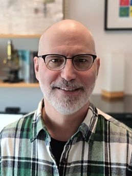
We are pleased to welcome Qian Lin, who has joined us as Assistant Professor in CSB to work on systems neuroscience, at the intersection of computational biology and neurobiology. In her experiments, she observes neurons light up across the entire brain as zebrafish make decisions in order to understand how the brain generates complex behaviours. She observes that “The whole brain is involved in decision making, but where is the best place to start? I start by using the cerebellum as the entry point.”
Her work challenges assumptions of how decision-making and movement are related in the brain. The cerebellum was long viewed as being solely involved in motor control, but Prof Lin's work and others have shown that there is no strict dualism of cognition and behaviour. Preparatory activity in the cerebellum is predictive of a decision to turn left or right, and of signals to take action.
Professor Lin reflects on this phenomenon as she learns a new punch or kick when practicing martial arts. She senses that the decision and the movement become closely linked and reflects on how this process occurs in her own brain.
An international scholar comfortable in China, Austria or the States, Lin grew up in the countryside of Hubei province. She would devour all the books in her home, at her neighbours’ homes and in the small school library. When she looked up from the pages of her Nature books, Lin would observe the animals all around her and wonder “What are they thinking about?”
As the first one in her extended family to start university, Lin had no single interest and thought she might follow her family into teaching. However, a field trip to Yunnan province revealed the great variety in biology as she gazed at the giant water lilies in Xishuangbanna Tropical Botanical Garden. She then applied herself to studies in structural biology, computational biology and neuroscience.
Lin left China in 2011 to start her PhD at the National University of Singapore supported by a NGS scholarship. The transition was difficult due to the culture and the language barrier of a new country. Her scholarship was dependent on maintaining good marks, which added to the stress of the transition. She was grateful that her supervisor Suresh Jesuthasan and labmates didn’t let her sit in the corner by herself doing experiments but invited her to join them and supported her studies.
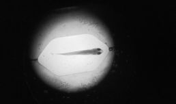
Lin’s PhD lab used zebrafish as a model system to study sensory representation and behaviour. The transparent bodies of live zebrafish larvae allowed them to view the entire nervous system. Lin found herself imagining complex behaviour experiments while she studied the neurobiology of light processing.
“I find neural circuits fascinating as you can get close to understanding underlying neural mechanisms”, says Lin. “By acquiring quantitative results, you approach the idea of predicting what zebrafish will do. Since you can see each cell within the transparent body, you can target specific regions of the brain to change their behaviour.”
Lin chose Alipasha Vaziri’s lab in Vienna for her post-doctoral research using new optical neurotechnologies to study complex behaviours. Early days in the lab were a struggle, with no results from her zebrafish training during the first six months. There was added difficulty as the lab moved to New York. Over time and with perseverance, her results poured in and careful analysis resulted in a publication in the prestigious journal Cell: “Cerebellar Neurodynamics Predict Decision Timing and Outcome on the Single-Trial Level”.
Lin acknowledges that being a scientist can be emotionally draining due to the various challenges it entails. For her, the stress of working as a post-doc with a toddler while seeking a faculty position during the pandemic stands out. Lin emphasizes that it’s important that you don’t give up. She finds it best to relieve tension through physical activity like martial arts and finding challenges that keep you mentally sharp. Lin advises that as things become more difficult, you develop the competency to deal with them and at some point you won’t have to relive how hard it used to be.
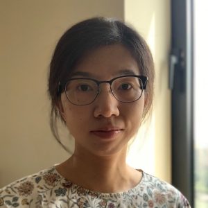 “My best decision was choosing an academic career,” Lin reveals. “With a PhD in neuroscience, I could have chosen to join industry as a data scientist or to continue in academia as a post-doc or professor. I chose academia because doing research is what I enjoy most.”
If you were to join Prof Lin’s lab as a student or post-doc, research would require some tool building of, for example, behavioural rigs, and would then occur on a weekly cycle: she prepares fish, images their brains and behaviour over 3 whole days, with two days for data analysis using code written in MatLab or Python. This all leads to a visualization of the process over time.
We are grateful that Prof Lin has chosen the Department of Cell & Systems Biology for her academic career. “I find Toronto to be chilly, but it is pleasantly quiet compared to New York. The neighbourhoods are very family friendly, with lots of nature and wildlife in the parks.” notes Lin. “My colleagues here have been generous with their protocols and with sharing their grant applications to help me to get my own funding.” We look forward to having her in the Department. Welcome!
Discoveries and Discussions at CSB Research Day
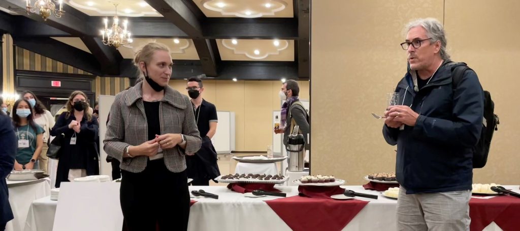
CSB held our Research Day for the first time in 2 years on September 22nd, 2022. Over 180 students, staff and faculty gathered at the Old Mill to hear the latest advances in research in the department. In his opening remarks, Graduate Associate Chair John Peever remarked how glad he was to see everyone in 3D after years of Zoom screens.
Our Keynote Speaker Prof Teresa Lee wowed the crowd with her detailed work on the epigenetics of longevity in C elegans. It was a pleasure to host her.
We are grateful to our alumni speakers who shared their career paths with our students. Declan Ali related fond stories of the Lange lab that led to his position as Chair of Biological Sciences at the University of Alberta. Gazzarrini lab graduate Carina Carianopol shared her more recent path to industry at Platform Genetics and related the joys of living in Niagara.
Trainee presentations were judged in different formats by faculty, post-docs or by applause. In addition to cash awards, some outstanding presenters also received prizes donated by Millipore-Sigma.
Oral Presentations
Senior students presented the details of their research in oral presentations with faculty scoring their presentations.
Jasmin Patel of the Gazzarrini lab at UTSC scored the highest marks for her talk on “Transcriptional regulation of FUSCA3 during seed coat development in Arabidopsis thaliana”
Our next award winner was Clare Breit-McNally of the Desveaux and Guttman labs at UTSG, who spoke on “The effector-triggered immunity landscapes of two Brassicaceous oilseed crops”
The final award went to “Spalt and Disco Define the Dorsal-Ventral Axis of the Developing Drosophila Medulla” from Priscilla Valentino of the Erclik lab at UTM. For more details on this research, you can read her brand new publication in the journal Genetics.
Lightning Talks
Our Lightning Talks event gave students three minutes to introduce their topic and to relate their findings. These rapid-fire talks were judged by the enthusiasm of the audience response.
Vineeth Andisseryparambil Raveendran from the Woodin lab was awarded the prize for his Lightning Talk on “Enhancing potassium-chloride co-transporter-2 (KCC2) function in neurons by targeting protein-protein interactions”. As the presenter who received the loudest applause, he was presented with some additional Millipore-branded gifts.
Ernest Iu, Milena Russo, Gregor McEdwards and Kailynn MacGillivray also won awards for the enthusiastic waves of applause inspired by their talks.
Poster Session
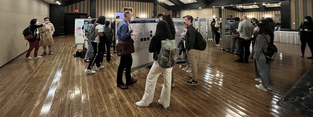
While attendees sampled canapes and drinks from the bar, mid-PhD students gave poster presentations over two sessions that were judged by faculty and post-doctoral researchers.
Samiha Benrabaa received the highest scores of all the poster winners, earning a special selection of Millipore-donated goods for her poster on “Ecdysteroid-signaling and reproduction in Rhodnius prolixus, a vector of Chagas disease”.
Russell Luke, Samuel Delage and Tamar Av-Shalom also earned awards for their excellent poster presentations.
As the day wrapped up, CSB chair Nick Provart gave his warm thanks to the organizing committee and its leader, Madison Marshall for a flawless day.

