
Composite biomaterial conveys tension across the wrinkled Drosophila embryo surface
Rowan Naidoo has uncovered the mechanism that allows plasma membrane architecture and the actomyosin cytoskeleton to work together to generate tension during early Drosophila fruit fly development.
He has published this part of his PhD work in the Harris lab in Journal of Cell Biology as “Actomyosin cortex integration with complex plasma membrane topography in the early Drosophila embryo.”
Naidoo co-authored the paper with Rebecca Tam, now a postdoctoral researcher at Rockefeller University, whose previous work from the lab explored how the walls between nuclear chambers form in the multinucleated Drosophila embryo.
Naidoo was intrigued by the fact that the function of the complex membrane topography that exists before these chambers arise remained unclear.
Understanding complex folds
The early Drosophila embryo has a plasma membrane that is highly folded; Naidoo’s work reveals how actomyosin networks integrate with these folds to generate tension across the embryo surface.
“This work really contrasts with what I learned from the textbook as an undergraduate,” notes Naidoo. “That depiction of the plasma membrane was as a flat sheet, with all the material underneath.”
“With my first assay with a lipid probe, I looked under the microscope and the plasma membrane looked extremely complex and hard to connect to the textbook image.”
Surface tension is important for the physical integrity of embryos, but materials typically fold because of compression. Thus, it was unclear how actomyosin networks generate embryo surface tension across a highly folded plasma membrane.
Naidoo’s research provided evidence of actomyosin networks integrating throughout the folds to form a composite material that produces surface tension and indicates a role for the Arp2/3 protein complex that drives network structure.
A diversity of techniques unveils what happens at the folds
 Naidoo’s research led him to master a number of techniques as he studied early stage Drosophila embryos.
To determine whether the folded surface was under tension, Naidoo used laser ablation to cut the membrane and measured membrane recoil from the wound. When myosin levels were low, membrane recoil was substantially decreased, indicating that actomyosin networks generate tension across the folded embryo surface.
Genetic perturbations and drug treatments demonstrated that myosin activity is indeed required for generating the surface tension, and that actin filaments interconnect the composite material.
Using dual live-cell imaging, Naidoo visualized the plasma membrane alongside cytoskeletal molecules in living embryos. He observed that plasma membrane folds condense and expand during cycles of embryo growth, while cortical myosin accumulates and dissipates in synchrony with these changes.
He also revealed that cyclic condensations are preceded by periods of expanded spacing between plasma membrane infoldings driven by the proteins of the Arp2/3 network.
Overall, these techniques provided evidence of the membrane architecture and actomyosin cytoskeleton forming a composite material that generates and transmits force across the embryo cortex. This surface tension is crucial for development of the Drosophila embryo.
The study expands understanding of the early embryo surface.
When viewed together, Naidoo’s findings reveal that plasma membrane folds act as a physical template for actomyosin assembly and activity. Additionally, distinct Arp2/3-dependent actin networks expand through the folds, and regulate their association with actomyosin networks.
The findings suggest that plasma membrane topography plays a fundamental role in the generation of cortical tension during development. Early mammalian embryos also rely on myosin-based surface tension and exhibit plasma membrane folding so this advance has potential relevance to screening in assisted reproductive technologies.
The deep folds of the plasma membrane imply that there could be interactions with other membranes within the cell. The team is now interested in looking at how these folds might also engage the endoplasmic reticulum found below the embryo surface.
Congratulations on this impressive insight into the biophysics of the early embryo!

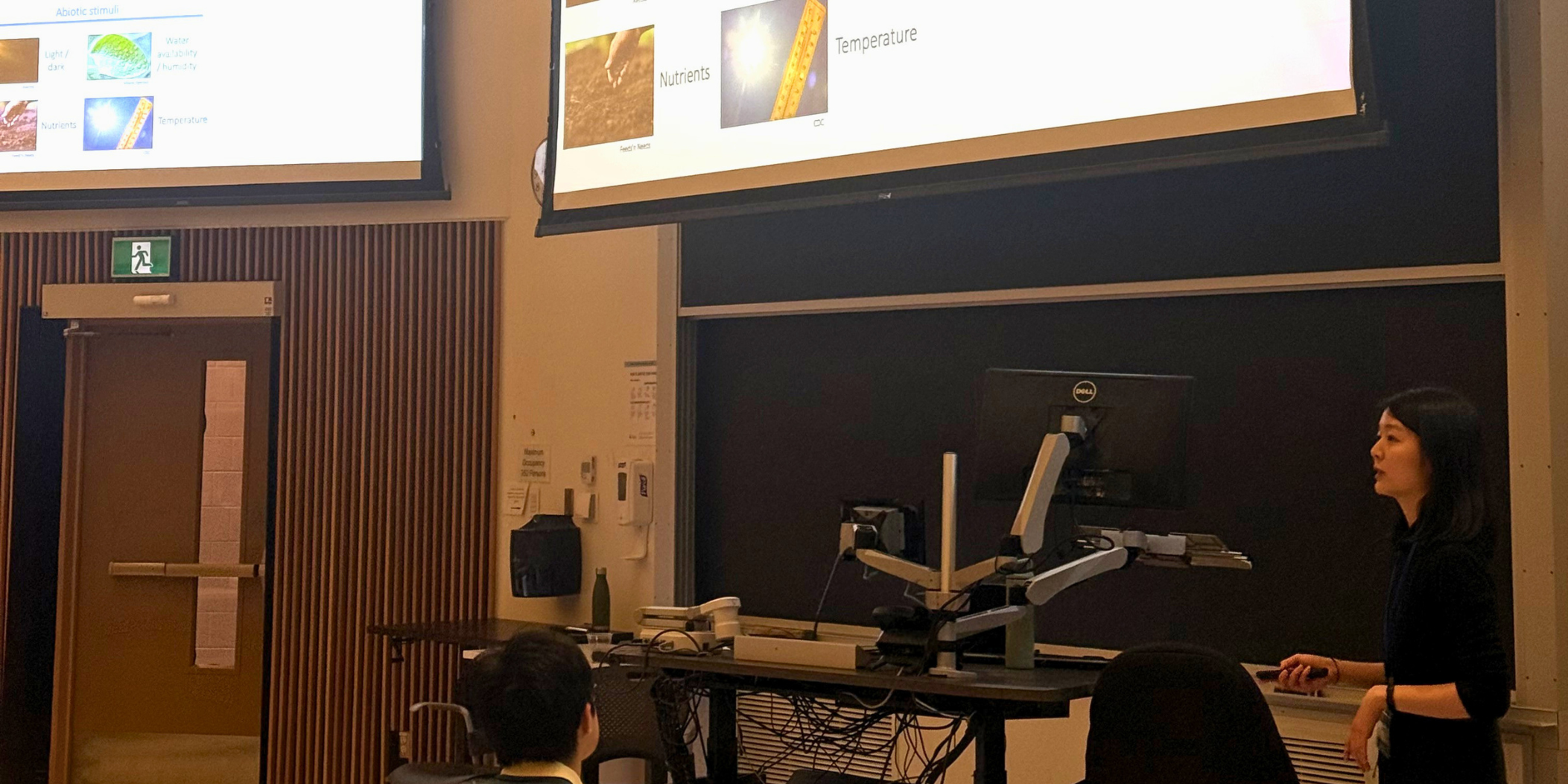
Drought response requires fine-tuning of RNA processing revealed by new research from the Provart lab
Subtle alterations in RNA splicing fine-tune how genes regulate drought tolerance in plants according to new research from Hasna Khan in the Provart lab. This exposes a new way to understand drought response that complements the usual focus on gene expression levels. These findings are published in The Plant Journal as "Differential splicing fine-tunes guard cell gene expression and is required for drought tolerance in Arabidopsis thaliana"
Revealing another layer of regulation
Professor Nicholas Provart studies how plants respond to environmental stress. As climate change increases the frequency and severity of drought, understanding how plants conserve water has become more important.
While many studies focus on which genes become active during drought, Provart and PhD student Hasna Khan suspected another layer of regulation might be hidden within guard cells, the specialized cells that control tiny pores on leaf surfaces.
“We started asking whether we could look at whether expressed gene transcripts were spliced into distinct mRNAs within the same cell”, Khan recalls. “I don't think either of us expected it to become as complex a project as it did.”
Guard cell-specific changes in drought response
Guard cells play a critical role in drought response. By opening and closing stomata, they regulate both carbon dioxide uptake and water loss. “Drought happens slowly as rains falter and soil dries,” explains Provart. “We anticipated fine-tuning in gene activity in guard cells as drought progresses, in addition to the more widely studied fast responses.”
A previous student in the Provart lab, Dr. Anna van Weringh, spent years developing methods to isolate RNA specifically from guard cells to test this hypothesis. The work presented substantial technical challenges because guard cells represent only a small fraction of the cells within a leaf.
“Getting enough guard cells that you could do RNA work, while also working quickly enough to avoid RNA degradation, was a really fine balance,” Khan says. “At times, it felt like I was elbows deep in guard cell RNA,” she adds with a smile.
Whereas van Weringh focused on changes in gene expression in guard cells from drought-affected plants, Khan analyzed the RNA-seq data to identify different splicing patterns using multiple bioinformatic pipelines. She then validated these results experimentally.
Khan discovered widespread changes in alternative splicing as drought progressed. Many of these changes were specific to guard cells and could not be detected when examining whole-leaf tissue.
“That is really one of our coolest findings,” says Khan. “When we see these guard-cell specific changes, it highlights how import it is to focus on this cell type.”
Drought-responsive alternative splicing required for optimal drought response
This insight suggest that alternative splicing acts as a second layer of drought-responsive gene regulation. While immediate drought responses rely on rapid signaling pathways, alternative splicing may allow plants to gradually adjust their physiology as drought conditions worsen.
Among dozens of candidate genes, SAFE1 emerged as particularly intriguing. This gene reproducibly showed drought-responsive alternative splicing in guard cells but maintained constant gene expression. This allowed the researchers to isolate the effect of splicing from changes in gene activity.
In an impressive confirmation of its importance, when SAFE1 was locked into one splice configuration the plants were more drought sensitive. “That really drove home the idea that this drought-responsive alternative splicing is required for an optimal drought response,” Khan says.
Avenues for new research
This discovery raises new questions about why plants use alternative splicing to regulate drought responses. One possibility is that alternative splicing provides a way to fine-tune the activity of genes with multiple roles throughout the plant, allowing guard cells to respond to drought without affecting other tissues.
Building on these findings, Khan’s next steps will focus on investigating drought-responsive alternative splicing in tomato guard cells. By exploring whether similar mechanisms exist in crop species, this research could help scientists better understand how plants adapt to water stress and potentially inform future strategies for improving drought resilience in agriculture.

CSB teaching assistants recognized for guiding their students to excellence
The CSB TA Teaching Excellence Award recognizes the significant role of teaching assistants in the Department of Cell and Systems Biology and their key contributions to the learning experience of students. Selection is based on demonstrated excellence in teaching through innovation, enthusiasm, organization and effective communication.
TAs are nominated by their students; this year's recipients are very grateful for the support from students in their courses.
For Koji Hartley, it was his first time TAing BIO130. "Having enthusiastic students made TAing for the first time a fun learning experience for me. As someone who wants to pursue teaching in the future, I am so grateful for this acknowledgement."
Madison Fedele notes that "One of the most rewarding parts of teaching BIO130 was getting to support students as they developed confidence in the lab and connected course concepts to hands-on experiments. I really valued creating a welcoming and supportive learning environment. Being acknowledged by the students makes the experience especially meaningful."
Tara McDonnell revealed that “Its always a pleasure to TA CSB349. Even after several years, my students never cease to impress me with their perspectives and ideas throughout the course."
For Brittany Dugan, this was her final year as a TA "This award means so much to me! I have TAed CSB325 for my whole PhD, and I am so honoured by this acknowledgement in my final year. I'm really grateful that my love for this course could shine through, and so happy that it resonated with my students. You're all the best! Thank you for believing in me. I'm so glad I could leave an impact on you all."
Congratulations to these accomplished educators!

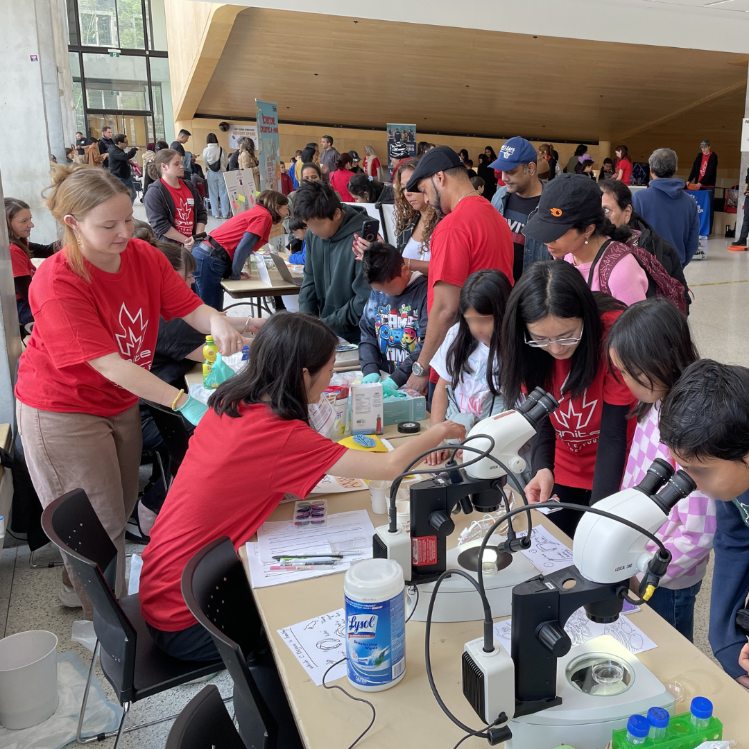
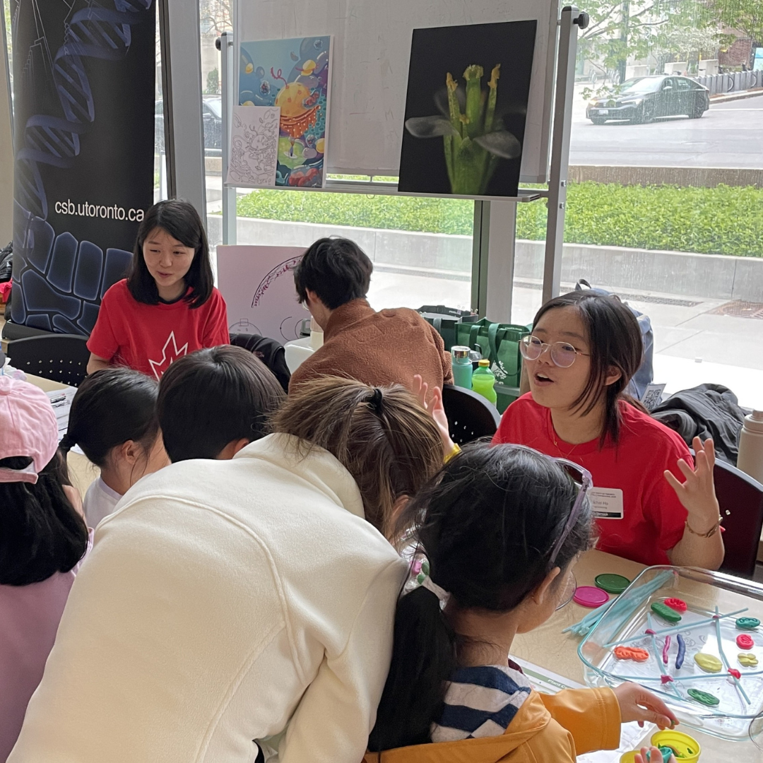
CSB shares research insights across life sciences at Science Rendezvous 2026
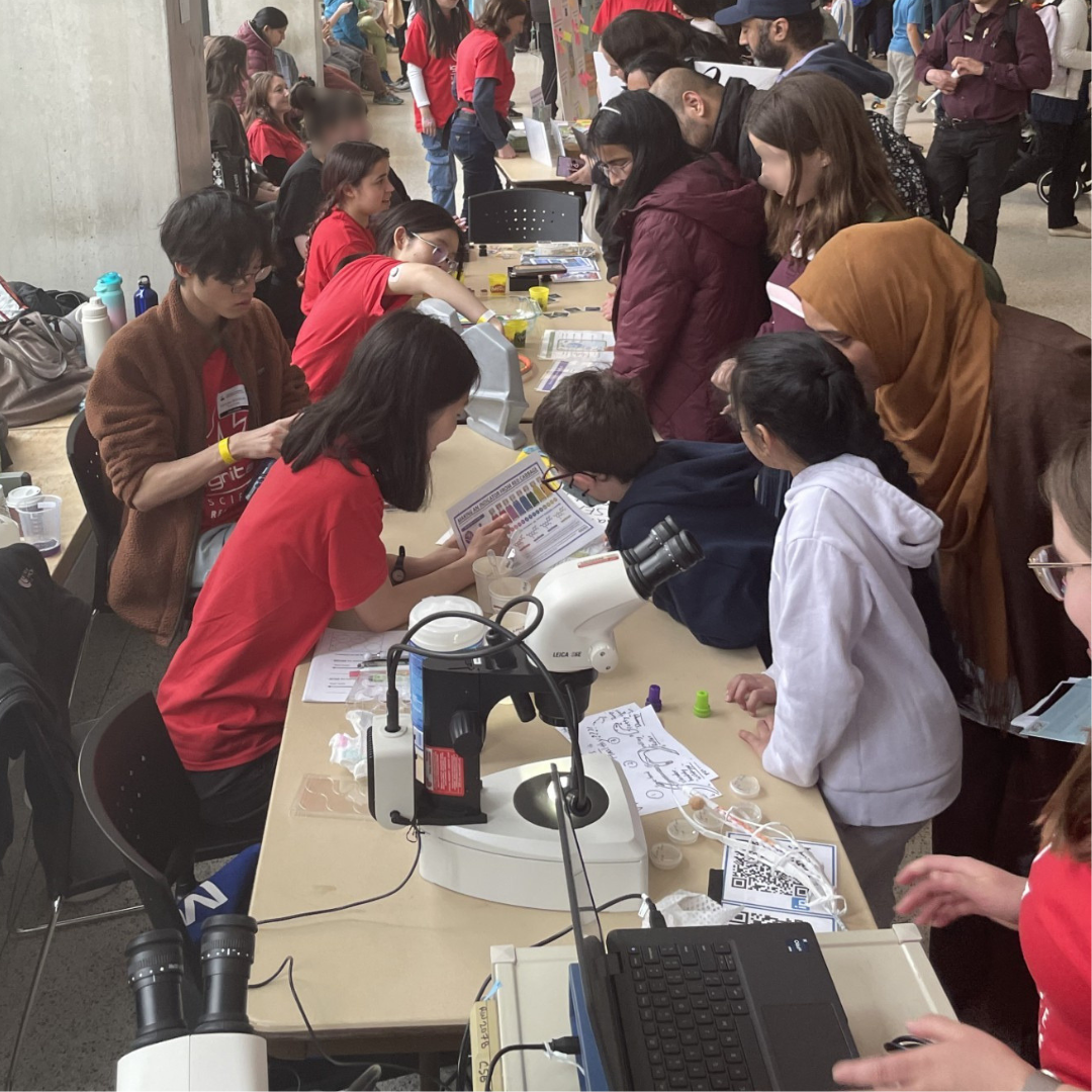
The diversity of life sciences research in Cell & Systems Biology was shared with the public through hands-on demonstrations at the Science Rendezvous science festival on a sunny Saturday, May 9th. Visitors explored neuroscience, developmental biology, plant biology, genomics, and cell biology while speaking with students, staff and faculty behind the research.
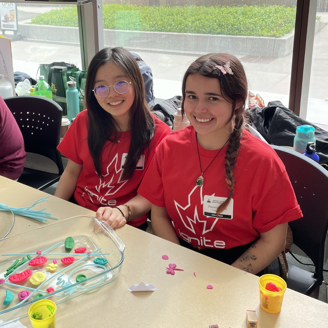
We were grateful for volunteers Rachel Ho, Phoenix Hewko and Nori Watanabe who learned quickly and who swiftly adapted to presenting several different activities.
Seeing life at the microscopic level
Visitors took home smartphone images of microscopic cells as Professor Heather McFarlane showed the fascinating shapes that cells take from discs to columns to jigsaw pieces. This demonstrations connected into McFarlane's research on plant cell wall biosynthesis and provided context for our Molecular Networks of the Cell specialization.
Neil Macpherson showed what was happening inside the cell by inviting visitors to view models depicting the organelles of plant and animal cells. They were encouraged to use pipe cleaners and modelling clay to create their own chloroplasts, mitochondria or other components of Cell & Molecular Biology inside glass dishes representing the cell.
The way cells work together to build a body was shown by regenerating planarian flatworms. After decapitation, they could regrow one head...or two heads(!) depending on chemical signals studied in our Stem Cells & Developmental Biology specialization. This wonky worms exhibit was developed by Professor Ritu Sarpal and demonstrated by Edlin Liang.
Kitchen science reveals surprising links to modern research
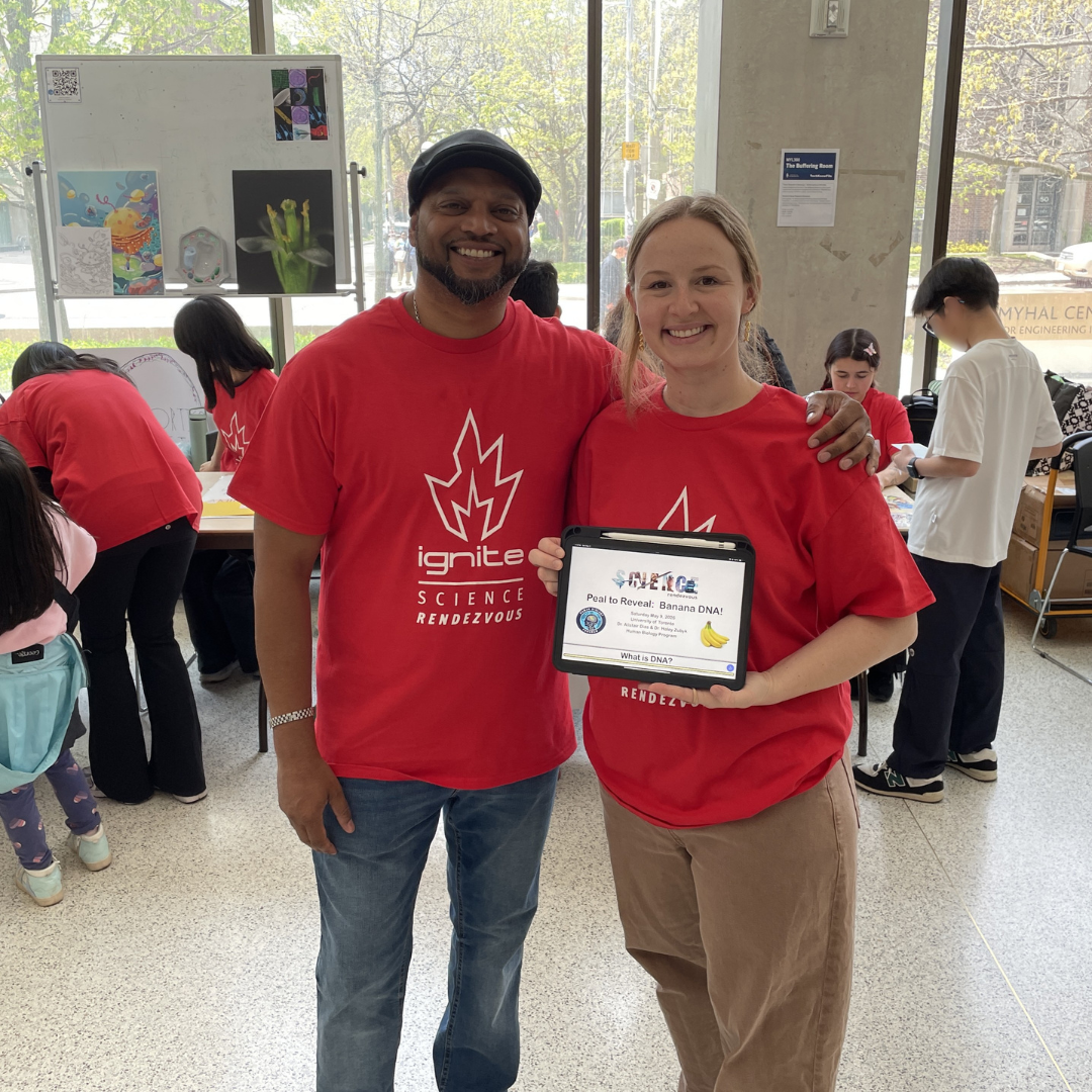
Visitors squished bananas to break them apart and used kitchen supplies to extract DNA as Human Biology Professor Alistair Dias and Professor Haley Zubyk described how this 'Peal to Reveal' technique worked to reveal stringy white strands of DNA. They went on to explain how extracting DNA from prehistoric animal remains could guide genome editing using Genome Biology.
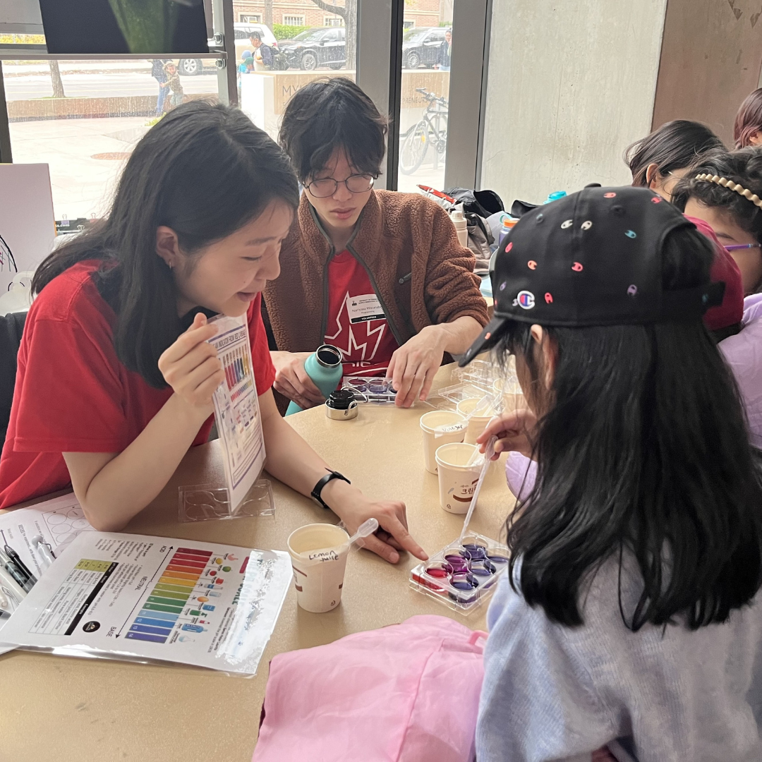
Curious guests observed colourful changes as red cabbage leaf extract responded to pH changes when they added soap, lemon juice or other liquids. Hyunsuh Lee explained how pH reflects levels of acid or base and described Plant Biology research in the Yoshioka lab looking at how pH and other environmental conditions affect plant immune responses.
Neuroscience across the animal kingdom
Visitors flexed their muscles as Stephanie Shisis demonstrated an electromyograph to measure their nerve impulses. This instrument is used by students in our Animal Physiology laboratories. As guests dotted with electrodes observed their own neurological traces, Shisis explained how she observes disrupted traces in her research on neurodegenerative disorders in mammals.
Wriggling worms showed neurological changes under the microscope as Ruby He demonstrated the C. elegans nematodes she studies as part of our Neuroscience graduate program. Since these transparent worms have only 302 neurons, He and the other researchers in the Calarco lab can trace the development of the nervous system through the life of the worm and look at the abnormalities that come from disrupted gene expression.
Showing off our beautiful science images
We presented colourful images depicting our research to the gathered crowds. There were lots of giveaways, as visitors took home flyers, stickers, banana DNA and a colouring page from artwork by Meghan Cao.
Thank you to all our presenters, and to Lisa Matchett, Reta Alam and Kenana al Kakouni who provided behind the scenes support!
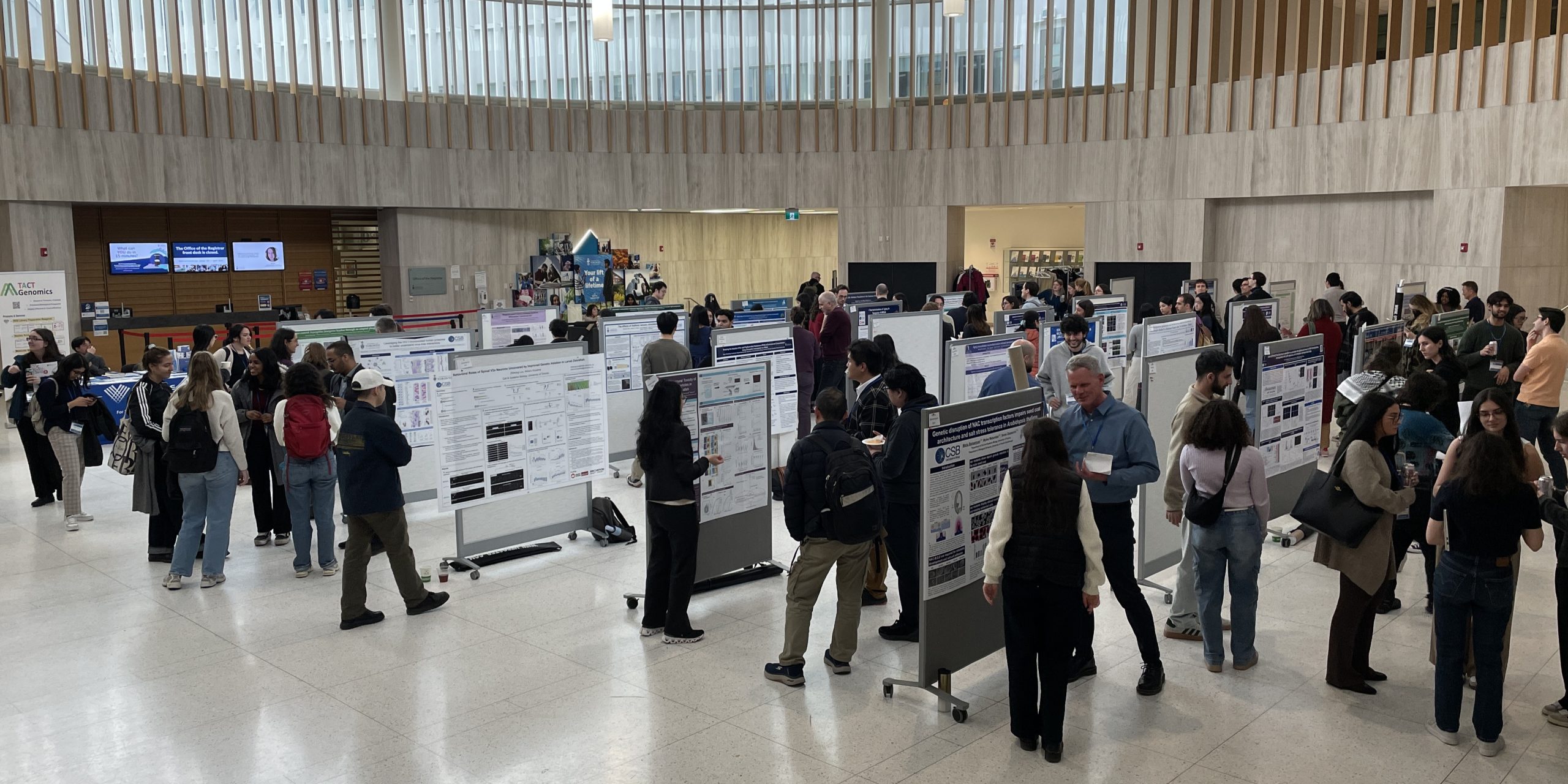
Research Day 2026 reveals astonishing advances from CSB across life sciences
CSB Research Day 2026 was held on May 1st at UTM's Kaneff Centre. This event showcased impressive innovation across the kingdoms of life.
Our keynote speaker, Professor Sheena Josselyn of SickKids Hospital presented her thought-provoking research on how memories are formed and erased in a Taylor Swift-themed talk.
We are grateful to Ryan Ruan (BSF) and Dr Burton Lim (ROM) who provided important job perspectives in a lunchtime career panel.
Through talks in the KN137 lecture theatre and posters in the Rotunda, CSB graduate students clearly and concisely presented their discoveries. Some presentations were chosen by judges as outstanding efforts and earned awards at the end of the day, presented by N. Ross Stewart and Bauhua Liu of the Organizing Committee.
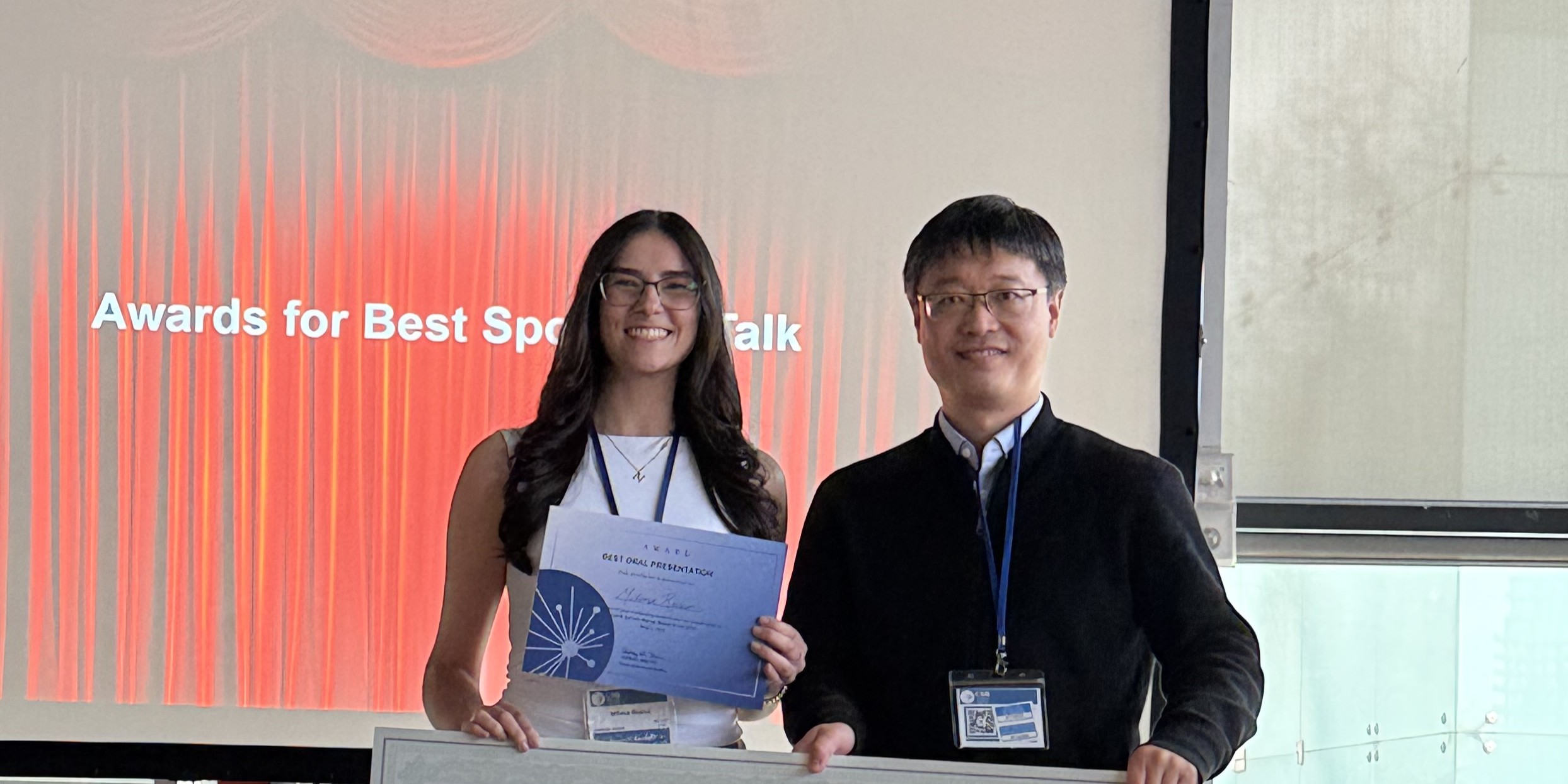
Best Spotlight Talk Awards
Claire Fernandes (Guzzo Lab) for "Emerging Cytometry Techniques To Purify And Study Virus Subpopulations"
Milena Russo (Liu Lab) for "Circuit Mechanisms Underlying Global Motion Processing in the Brainstem"
Best Lightning Talk Award
Anthony Kadamani (Peever Lab)
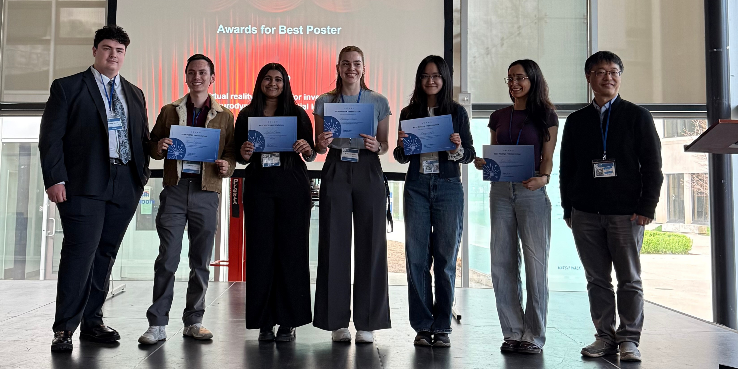
Best Poster Awards
Matthew Danesh (Peever Lab)
Congrong (Ruby) He (Calarco Lab / Zhen Lab)
Serene Moussaoui (Terebiznik Lab)
Ryan Ugovsek (Lin Lab)
Mackenzie Wilson (Phillips Lab)
Congratulations to these talented presenters! Thank you to the Organizing Committee N. Ross Stewart, Bryan Guo, Gary Chatha, Boahua Liu, Adam Mott, Shelly Lumba, Ben Eldridge and Denise Horsley. Photographs by Angela Sidsworth.


Epigenetic markers of neuronal gene regulation revealed to earn Hone-Buske award
Kailynn MacGillivray has earned the Dr Christine Hone-Buske Award for her outstanding publication “Widespread association of Polycomb complex–deposited histone H2A monoubiquitylation with enhancers and neuronal gene regulation”. This research in Science Advances reveals the role of protein modifications in the developing nervous system of the worm C. elegans.
MacGillivray’s work in the Saltzman lab focused on ‘epigenetic’ changes promoted by the protein complexes PRC1 and PRC2. These changes act on histone proteins bound to DNA, serving to turn genes off by packaging them tightly or turn them on by opening up the DNA surrounding the gene.
MacGillivray focused on ubiquitin and methyl molecules attached to histones. “We observed what we thought was a pretty unique phenotype in the worms if you look at PRC1 mutants for C. elegans versus PRC2 mutants.” MacGillivray explains.
“We found that genome-wide these histone modifications weren't working together in the same way as in mammals or fruit flies. Overall methyl patterns were unexpectedly maintained when ubiquitin was depleted.”
Given this unusual observation, MacGillivray used genome analysis to reveal that genes misregulated in ubiquitin-deficient mutants are enriched for nervous system functions including neuronal differentiation and axon guidance.
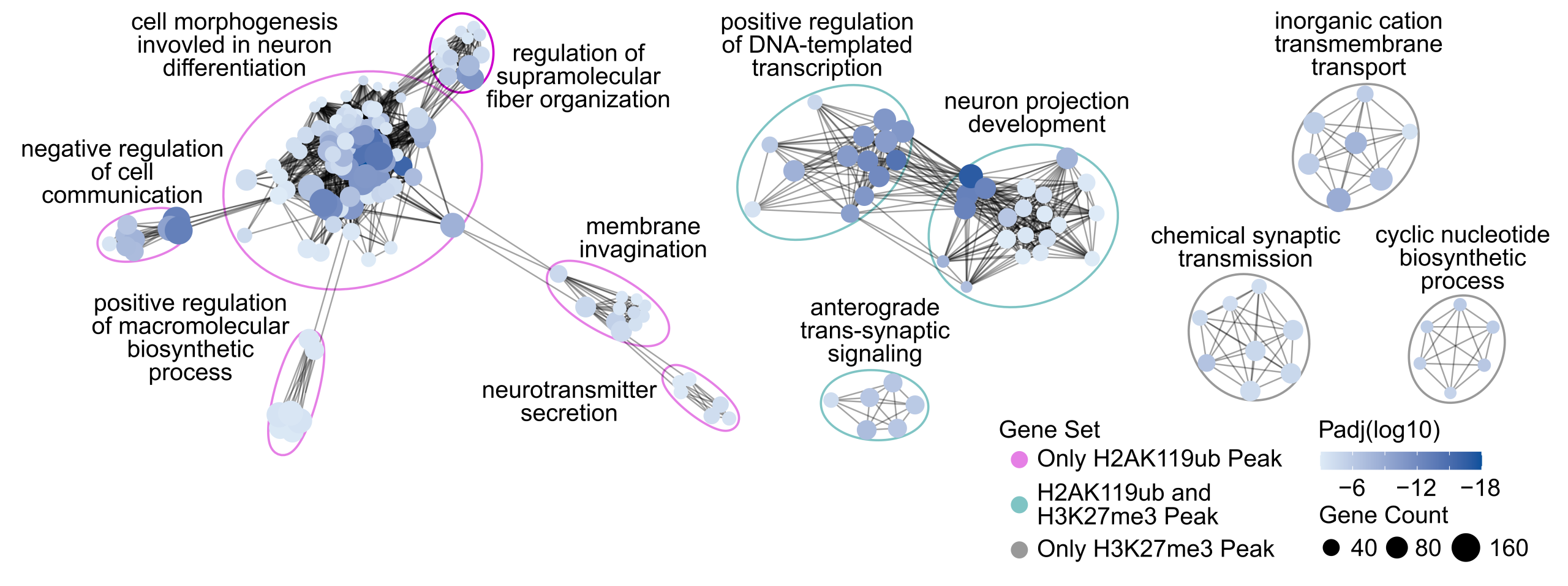
An exciting insight from this analysis was that epigenetic changes in PRC1 mutants were enriched at ‘enhancer’ regions: sequences outside of genes that regulate where and when a gene is expressed, even at a distance.
MacGillivray was puzzled: “We had this enrichment at enhancer regions and at neuronal genes, but we didn't have an obvious link in terms of a gene regulatory standpoint how these two are related in the developing worm. Then we were looking through recent papers and saw a set of enhancers that become nicely accessible or potentially active within neuronal tissue.”
“I found that ubiquitination was most highly enriched at these enhancers compared to enhancers that are accessible in other tissues,” MacGillivray explains. “That was definitely the ‘oh, this makes sense!’ moment that tied this paper together.”
There was excitement in the Saltzman lab as MacGillivray showed the results to her colleagues and mentor. Their animated discussions postulated that epigenetic changes drive temporal control as the nervous system develops and this may be what is constraining activity.
The unique aspects of worm neuronal development can provide insights into other organisms. Some neurodevelopmental dysfunctions in humans are linked the PRC1 complex, and the Saltzman lab is collaborating with researchers in Miami to see if worms provide a model for understanding these changes in humans.
“It’s been great to follow all these different pieces of evidence,” says MacGillivray. “I’ve always been curious, and making these discoveries to me is the whole point of science. I’m glad to have this enthusiasm rewarded with the Dr Christine Hone-Buske Award.”
Congratulations, Kailynn!

Intensive Laboratory Experience prepares Life Sciences students for a career in research
The Human Biology Lab Bootcamp is an intensive two-week wet and dry lab training program offered to students finishing their third or fourth year of studies in life sciences. It provides hands-on research experience, technical skill development, and guidance for students considering careers in biomedical research or related fields.
The HMB Program will host the 2026 Lab Bootcamp on May 4–15. This no-cost, ungraded program is designed for students with fewer opportunities to gain hands-on biology lab experience. The Bootcamp was conceived and designed in 2017 by University President Dr Melanie Woodin (then Director of HMB) by Dr Colleen Dockstader.
Unlike weekly lab classes, each day’s experiments build on the previous day’s work—like chapters of a story—so students follow multi-day workflows. Topics include DNA purification, restriction digestion, cloning, gel extraction, ligation, colony PCR, tissue culture and transfection, immunofluorescence microscopy, and more.
Groups of eight students are supported by one facilitator. Those accepted for the program are always looking to maximize their skills and knowledge without the stress of regular tests.
The program gives special recognition to Drs Colleen Dockstader, Jasty Singh, and Alan Wong for creating the lab manuals and designing this Bootcamp in 2017.
As this program progressed, professors and postdocs helped sustain the program, including Drs Jessica Pressey, Naijin Li, and Samuel Delage, as well as many facilitators who taught students and guided them through the course each day.
We look forward to welcoming this year's cohort of dedicated students!

Prof Haley Zubyk Earns ASSU Award for Excellence in Teaching
Congratulations to Professor Haley Zubyk, who was nominated by Arts & Science students to receive the Ranjini (Rini) Ghosh Award for her Excellence in Teaching!
The award was earned by Zubyk’s ability to stimulate and challenges students’ intellectual capacity, skill at communicating the course material to students, mastery of the subject area and for being highly accessible to students
"Receiving this student-nominated award is incredibly meaningful to me because it reflects that my students feel supported throughout their undergraduate journey and that my passion for teaching is visible to them,” Zubyk says. “Teaching and learning are deeply rewarding, and my students are without question the best part of this work.”
Zubyk's students experience her passionate pedagogy studying the Challenges of Antimicrobial Resistance in Human Biology and attending her course on AIDS: A Global Perspective in the Fall term and in Summer sessions. In the Winter term, Zubyk presents an Introduction to Human Biology and shares insights into The Human Microbiome in Health & Disease.
Congratulations, Professor Zubyk!

A year of deep research leads to a culminating event for fourth year students
Fourth year students in CSB can join our research laboratories to conduct projects in their favourite topic as part of CSB497, 498 or 499 courses. Their final task after generating data and analyzing results is to present their conclusions at our annual Undergraduate Poster Session.
This year, the poster session was held on March 27th and featured dozens of posters showing students' discoveries in cell biology, cancer biology, developmental biology, neurobiology and plant biology.
The posters were judged by departmental staff and faculty. They assessed the content of the posters and the presenters' ability to describe their projects and answer questions.
The F Michael Barrett Award was presented to students who excelled at their presentation by Undergraduate Chair Professor Keiko Yoshioka.
Faten Abla (Bruce lab) showed their expertise in "Investigating the Role of Eph/Ephrin Signaling in Tissue State Transitions During Zebrafish Mesoderm Internalization"
Maia Edney (Yip Lab) pursued "Investigation of Novel Compound for Suppression of Extracellular Matrix Proteins"
Alex Huang (Plotnikov Lab) demonstrated how "ER Ca2+ Regulates ER: PM Contacts to Facilitate Linear Migration in Elevated ECF Viscosity"
Tiantian Lei (Lin Lab) studied the "Effects of GCaMP6 on Effects of GCaMP6 on the Survival and Behaviour of Zebrafish Larvae"
Aliya Mohd Zamri (McFarlane Lab) took on the task of "Confirming putative cell wall signaling mutants from a forward genetic screen"
Shannon O’Reilly (Calarco Lab) demonstrated a "High-Throughput Behavioural Monitoring Platform to Identify RNA-Binding Proteins Regulating Locomotion in C. elegans"
Zoe Smith (Yip Lab) presented results from "Investigating the Role of FOXO1 in Radiation-Induced Fibrosis"
Tea Reed Watson (Lumba Lab) showed how "ShHTL7 mutants demonstrate altered germination and temperature sensitivity compared to wild-type in an Arabidopsis model"
Fiona Yi Yang Xu (Bruce Lab) explored "Frmd4 Proteins at Cell Junctions: Potential Cytohesin Adaptors during Zebrafish Epiboly"
Vicky Ziyi Wei performed her work in the Tepass lab, which studies the cellular and molecular mechanisms that control the polarity of epithelial cells and the cell adhesion between epithelial cells that binds cells into tissues
Congratulations to these impressive students on their accomplishments and good luck as you move on to graduation!